


Introduction
Plants are a rich source of bioactive metabolites and phytochemicals, which are used for their medicinal and therapeutic characteristics [1]. Due to this characteristic, humans have been using plant parts to treat various diseases since time immemorial and are considered an alternative healthcare medicine for approximately 85% of the world's population [2,3]. Plants with medicinal properties commonly contain bioactive compounds such as tannins, saponins, flavonoids, alkaloids, steroids, and other derivatives [4,5,6]. Nowadays, these bioactive compounds are studied thoroughly to determine their physiological, behavioral, and immunological effects, which can be beneficial to humans.
Despite the use of medicinal plants to treat various diseases, there is still a lack of valid experimental evidence for their adverse effects [7]. Depending on their origin and nature, medicinal plants can be a source of toxic element exposure. For instance, preparation and use of medicinal plants can cause damage to human health [8]. Numerous studies have already been conducted to investigate the toxicity of medicinal plants on human organs such as the kidneys, heart, liver, and skin [9,10,11]. Studies have found that certain plants contain toxic elements such as arsenic, which causes skin pigmentation, skin cancer, and hyperkeratosis in feet and hands [12,8,6]. Plant extract toxicity can also be fatal due to poisoning manifested as vomiting, lightheadedness, and heart block [13]. Olayode et al., [14] showed that the repeated treatment of leaf extract of Stachytarpheta cayennensis (blue snakeweed) to male and female rats led to hemorrhage, pyknotic nuclei, vascular congestion, tissue necrosis, and disruption of hepatocytes (liver) arrangement. In addition, Chebaibi et al., [15] also showed that the use of plants traditionally used in the treatment of edema and renal colic is toxic to organs of the body, such as the liver. Aside from that, insufficient research on herbal medicines with uncertain pharmacologic action poses a danger to developing fetuses [16,17]. The most documented adverse effects of medicinal treatments during pregnancy include uterine contraction stimulation and abortion [18]. Various traditional medicines have ingredients that can cross the placenta and reach and impact the fetus, and certain natural medicines have been documented to cause pregnancy loss due to hormone imbalance that could end in abortion [19,20]. With these reports, the World Health Organization (WHO) emphasized the safety of herbal medicines by issuing guidelines for herbal medicine assessment and emphasizing the importance of toxicity studies [7].
One of the commonly used medicinal plants from the Philippines is Decalobanthus peltatus, locally called Buwakan, from the Convolvulaceae family in the Philippines. It is a coarse, woody vine with alternate, smooth, and somewhat rounded leaves [21]. Extracts from these plants are used to treat diarrhea, cough, abdominal pain, sore eyes, and wound inflammation and aid during childbirth [22]. Studies have shown that it has antioxidant and alpha-glucosidase inhibitor activities, which can be helpful in diabetic treatment [23]. This plant is also reported to contain bioactive compounds with broad antimicrobial activity [21]. Despite the usefulness of this plant extract, there are limited reports regarding the embryotoxicity and teratogenic effects of D. peltatus extracts. To address this need, this study investigated the embryotoxic and teratogenic effects of D. peltatus crude leaf extracts on duck embryos [24, 25, 26]. Cost-efficient extraction of plant materials was achieved by employing the maceration technique with methanol as the initial extraction solvent, followed by subsequent liquid-liquid extraction with water, n-hexane, and ethyl acetate [27, 28]. To the best of our knowledge, this will be the first report in the Philippines evaluating the toxic effects of D. peltatus in animal models.
Materials and Methods
Study area
The plant samples were collected from the municipality of Cagdianao, Dinagat Islands (Fig. 1), where it is used as medicine such as wound healing by the local inhabitants. A gratuitous permit was provided by the Department of Natural Resources, allowing the researcher to collect samples from Dinagat Island, Philippines.
Morphological and molecular identification of the plant
The plant samples were identified and confirmed by a Botanist from the Department of Biological Sciences, College of Science and Mathematics, MSU-Iligan Institute of Technology, Iligan City, Philippines. Voucher specimens (USTH 017-729) were deposited at the University of Santo Tomas Herbarium.
To further confirm species identification, DNA extraction was performed in the Developmental Biology laboratory, PRISM, MSU-IIT, Iligan City, Philippines, and was done following the protocol provided in the DNA extraction kit (Invitrogen Life Technologies, USA).
The PCR amplification, purification, bidirectional DNA sequencing, and analysis of the DNA extracted from the sample were performed in Macrogen Incorporation (Seoul, South Korea). PCR amplification and DNA sequencing were carried out as per the protocol developed by the manufacturer. The primers used are the forward primer: ITS3- 5’GCATCGATGAAGAACGCAGC3’ and Reverse primer: ITS4- 3’TCCTCCGCTTCTTGATATGC5’, targeting the internal transcribed spacer 2 (ITS2) region of the ITS sequence.
Methanolic Crude Extract Preparation
The plant pieces were air-dried in the shade for two to three weeks until all moisture had been eliminated. The dried leaves were finely macerated and then immersed in 100% absolute methanol at a 1 g : 4 mL ratio. For three days, the soaked plant pieces were stored in a dark spot. Then, the extracts were filtered using a Whatman no.1 filter paper. A rotary evaporator (Rotavapor® R-100, Buchi, Switzerland) was used to remove the extraction solvent and get the concentrated plant extracts [29, 30, 31]. Serial dilution for the crude extract was then performed.
Preliminary Phytochemical Screening
The crude extract of D. peltatus was subjected to preliminary phytochemical screening performed by a Registered Chemist from the Department of Chemistry of the Mindanao State University- Iligan Institute of Technology using standard methods [ 32, 33] with modifications.
Test for Alkaloid
2 mg of the crude extract from the prepared stock solution was added to 2 mL of 2 M of HCl. The filtrates were then treated with 1 mL of potassium bismuth iodide solution (Dragendorff's reagent). The formation of an orange precipitate determined the presence of an alkaloid.
Test for Flavonoids
A few drops of 1.0 M sodium hydroxide solution were added to 2 mg of the crude extract. The appearance of a strong yellow color that turns colorless upon the addition of HCl indicates the presence of flavonoids.
Test for Saponins
About 5 mg of plant extract was mixed with 5 mL of distilled water. The solution was shaken vigorously. Persistent frothing indicates the presence of saponin in the extract.
Preparation of Water, Hexane, and Ethyl Acetate Extract
After rotary evaporation, the concentrated plant extract was subjected to solvent partitioning. Three distinct solvents with various polarity indices were used for solvent-solvent partitioning: 100% hexane (0 polarities), 100 % ethyl acetate (4.4 polarities), and water (9 polarities). The partitioning was done in stages based on rising polarity [31].
Water and Hexane Partition
Partitioning was performed using the method described by the group of Abu (2017) with minor adjustments. One g of crude methanolic extract was dissolved in 20 ml of distilled water, then 20 ml of 100 % hexane was added. The hexane layers were filtered and concentrated under a vacuum in a rotary evaporator.
Water and Ethyl Acetate Partition
The hexane extract was partitioned further with ethyl acetate. The densities of the solvents determined the identities of the layers. The collected ethyl acetate layer was concentrated under vacuum in a rotary evaporator, and the water layer was freeze-dried and carefully packed in a container with parafilm. These samples were kept at 20 °C for the succeeding experiments.
Brine Shrimp Lethality Assay
Brine Shrimp Lethality Assay (BSLA) from an existing protocol with minor modifications [34, 35]. Five (5) g of brine shrimp eggs were placed in a brine incubator and immersed in 500 mL of synthetic seawater, with light and aeration, for 48 hours. The cytotoxicity test was carried out by weighing 50 mg of the extract, which was dissolved in 1 % of Dimethyl sulfoxide (DMSO) pipetted into concentrations of 1000 ppm, 500 ppm, 250 ppm, 125 ppm, 62.5 ppm inserted into vials. Three replicates were made for every concentration. Five mL of artificial seawater was added and homogenized with a vortex. Each vial contained ten brine shrimp larvae and 50 μL of a yeast solution as feeding. The vials were placed under the heat lamp and left for 24 hours. The LC50 value was calculated using the probit analysis of the Statistical Package for the Social Sciences (SPSS) application 26th Version. An LC50 value <1000 ppm is considered active.
Preparation of test organism
Fertile duck eggs (embryonic day 1) were acquired from a local vendor in Iligan City, Philippines. Dirt, excrement, and debris were removed from the eggshells and were placed in an aseptic egg incubator for a day for acclimatization. The candling method was conducted to check the viability of the eggs to be used and to check the location of the air sac and mark it. Eggs with underdeveloped and dead embryos were discarded.
Treatment of D. peltatus leaf extract on duck embryo
The desired concentration was obtained by dissolving 100 mg of crude extract into 10 ml of 1% DMSO and 100 mg of freeze-dried crude extract into distilled water as stock solution. The stock solution was serially diluted into five appropriate concentrations based on the result from the BSLA assay. For each concentration, 30 eggs were utilized, 10 for each day of observation (days 7, 14, and 21). Prior to treatment, the eggs were gently wiped with 70% ethanol once. A small hole was made around the air sac. The eggs were treated on day 3 of incubation. Then, the eggs were re-treated daily following the first treatment until the day of observation [36,37]. The treatments were injected through the hole using a sterile syringe. The holes were resealed with parafilm, and the treated eggs were incubated until observation. The process was done in a laminar flow hood to avoid contamination. This study employed a Completely Randomized Design (CRD), ensuring that all test solutions (D. peltatus leaf extract) and negative control were tested on duck embryos.
Analysis of the effects of D. peltatus leaf extract on vasculogenesis and angiogenesis
On day 7 of incubation, live embryos were selected for observation [38]. A 3x2 cm window was made on top of the embryo to view the vascular region or the chorioallantoic membrane (CAM) of the embryo. The vascular region was photographed and analyzed using an image analyzer software, ImageJ [39]. A specific region was extracted from the captured images. The extracted region was the right-lateral vitelline vascular plexus, measuring around 159 mm2 and containing 1100x1800 pixels [36]. The photos were converted to 8-bit format and treated to improve edge recognition in preparation for the next steps. Finally, their color levels were reduced to binarized (black and white) format, and the images were skeletonized. The skeletonized image depicts the object’s structural form. For each treatment and control group, ten (10) images were processed. The effects of D. peltatus on early embryonic vasculogenesis and angiogenesis were determined using the number of junctions, average branch length, vascular density, and vascular length density quantification.
Gross evaluation and measurement of the embryo
The embryos were examined at different time intervals of days 7, 14, and 21 of incubation [36]. At each time point, the eggs were opened, and the embryos were removed to examine any gross injuries or abnormalities on the body. The body weight was measured using a digital weighing scale. The average weight of the embryos was computed. At the end of each observation, all embryos were humanely killed by rapid cooling [40, 41].
Histopathological evaluation
Skin tissues from the duck embryos at days 7, 14, and 21 were dissected and placed in 10% neutral buffered formalin. Paraffin-embedded tissues were cut using a microtome at 6 μm thickness (HistoCore BIOCUT, Leica Biosystems). Serial sections were stained with H&E for investigation under a light microscope (Olympus CX22LED) [42,43].
Data analysis
All the graphs and statistical analyses were performed using GraphPad Prism software version 9.0 for Windows (GraphPad Software, San Diego, CA, USA). One-way analysis of variance (ANOVA) was conducted, followed by Tukey’s test to determine the significant difference between the ImageJ parameters among the embryos. Dunnett’s multiple comparisons test was used for diameter measurements of primary, secondary, and tertiary vessels. Two-way analysis of variance (ANOVA) followed by Tukey’s test was used to analyze the significant differences in the embryo weight of the experimental and control groups. A p-value of <0.05 was defined as statistically significant.
Results and Discussion
Phytochemical Screening of the crude extract of Decalobanthus peltatus
The phytochemical screening result yielded several bioactive compounds that include the presence of alkaloids, flavonoids, saponins, steroids, and tannins. Detected bioactive compounds are visualized by certain levels of abundance, where a + sign corresponds to the positive presence of the active compound, wherein a single, double, and triple + sign suggests the degree of detection in increasing order.
Table 1 enumerates the bioactive compounds found in the plant extracts and their level of detection. It shows the presence of alkaloids, flavonoids, saponins, steroids, and tannins.
The presence of these secondary metabolites in the leaf extract is significant as they may contribute to the traditional use of this plant to treat various ailments. Among these compounds, flavonoids, saponins, and tannins were abundant in D. peltatus. Substantial research into the health effects of flavonoids and their modes of action has gained the public's attention. Flavonoids are generally referred to as antioxidants since they are polyphenolic and may absorb free radicals [44]. While this feature may be valuable in food shelf-life research, it is exceedingly improbable that the health advantages of flavonoids are related to direct-acting antioxidant activity [45]. Saponins have several biological functions, including antibacterial, antifungal, antiviral, anti-inflammatory, anti-ulcer, hemolytic, and hepatoprotective effects [46, 47]. In addition, the saponins revealed the ability to operate synergistically with antibiotics, assisting in the recycling of old medications that were previously believed ineffective owing to resistance issues [48,49]. Tannins are the primary study topic in the development of natural alternatives to in-feed antibiotics due to their variety of biological functions such as antibacterial, anti-parasitic, antiviral, antioxidant, anti-inflammatory, and immunomodulation, among many others [50].
Brine Shrimp Lethality Test of the Different Crude Leaf Extracts
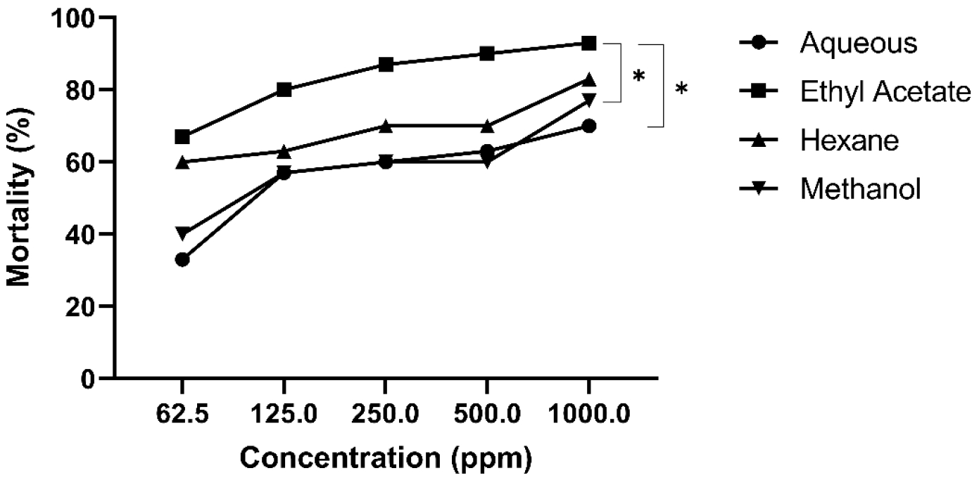
Initial investigations of the D. peltatus crude leaf extracts were done using the Brine Shrimp Lethality Test. This test provides results about the %mortality of Artemia salina exposed to five different concentrations of each crude plant extract (aqueous, methanolic, hexane, and ethyl acetate) from D. peltatus. The %mortality values were then transformed using the Probit Table, and the regression analysis was done with the log value of the concentrations to get their LC50 values (Fig. 1). The LC50 values are classified according to Clarkson’s Toxicity Criteria with LC50 value greater than 1000 ppm as non-toxic, 500-1000 ppm LC50 value as low toxic, 100-499 ppm as medium toxic, and 0-99 ppm as highly toxic [51]. Results of the study show a trend in which the higher the concentration of the plant extracts, the higher the %mortality values. Among the crude leaf extracts, the ethyl acetate of the D. peltatus shows a high % mortality after 24 hours, followed by hexane extract. Aqueous and methanolic crude leaf extracts exhibit similar %mortality (Fig. 1).
Table 2 shows the LC50 values of the different plant extracts with their corresponding toxicity level. Ethyl acetate and hexane leaf extracts of D. peltatus display LC50 values of 14.54 ppm and 26.30 ppm, respectively, which, based on Clarkson’s Toxicity Criteria, is considered highly toxic. The methanolic and aqueous fractions are considered medium toxic. Based on these results, the ethyl acetate extract, being the most toxic among the crude leaf extracts, was utilized to determine its effect during angiogenesis and during the development of the duck embryos.
The primary goal of assessing the safety of any medicinal plant is to determine the nature and significance of any detrimental effect, as well as the exposure level at which this effect is detected [52, 53, 54, 55, 56]. While there is a substantial phylogenetic gap between brine shrimp (A. salina) and the duck embryo, the use of brine shrimp as a preliminary assay is justifiable if it is viewed as a screening tool that helps prioritize compounds for further, more specific toxicity testing on vertebrate target organism (e.g., avian embryos). Ritieni et al. [57], for instance, conducted research on the toxicity of fusaproliferin (FP) and two of its derivatives to A. salina and tested their teratogenic effects on fertilized chicken eggs. In addition, Hlywka et al. [58] who assessed the toxicity of mycotoxin fumonisin B1 (FB1), reported that although the chicken embryo bioassay requires a higher concentration of toxin, the FB1's toxicity was found to be comparable between the chicken embryo and brine shrimp bioassay at a cellular level. Thus, this approach balances the need for safety assessment with the ethical considerations and resource constraints associated with animal testing [59].
The current investigation found that the degree of lethality was directly related to extract concentration. After 24 hours of monitoring, the highest mortality was recorded at 1000 ppm concentrations and the lowest mortality at 62.5 ppm concentrations. It was discovered that at higher concentrations of treatment extracts, the shrimps began to die after the initial hours of treatment, and by 24 hours, nearly all the shrimps had died. The presence of terpenoids, steroids, flavonoids, saponins, and fonolic compounds could be accounted for their cytotoxic properties [21,22]. Overall, the fraction with the highest %mortality is the ethyl acetate fraction, implying that the bioactive compounds of interest are of similar polarity with ethyl acetate. Different solvents may result in extracts with varying concentrations of bioactive compounds due to the varying solubility of these compounds in different solvents [29]. These differences in extraction yield may lead to differences in the dose-response relationship observed in the bioassay. In this case, ethyl acetate as an extraction solvent may have favored the extraction of polar compounds in higher concentrations, which might have influenced its toxicity. The ethyl acetate extract of D. peltatus was found to be the most toxic among all the partitions because it yielded the lowest LC50. This result shows that it only took 14.54 ppm of D. peltatus ethyl acetate extract to kill 50 % of the brine shrimps. The rest of the extracts needed a higher concentration. To observe how the toxicity of ethyl acetate would manifest as a teratogen, ethyl acetate extract was used for the embryotoxicity test on duck embryos.
Vascular branching pattern analysis
To elucidate the effects of D. peltatus during angiogenesis, chorioallantoic membrane assay (CAM) was utilized, and the vascular branching pattern was quantified in treated duck embryos as compared to control groups.
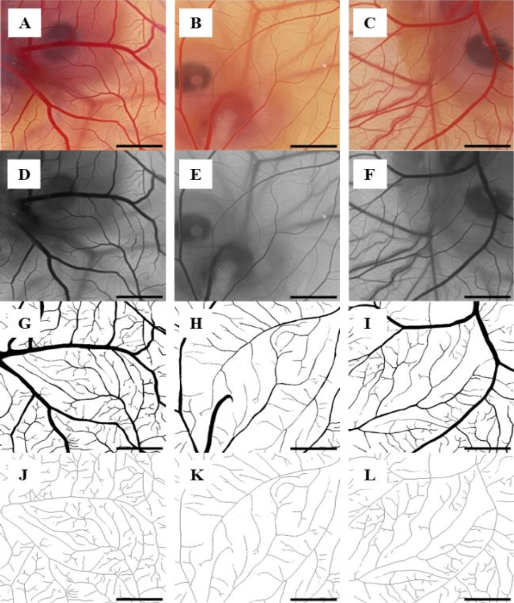
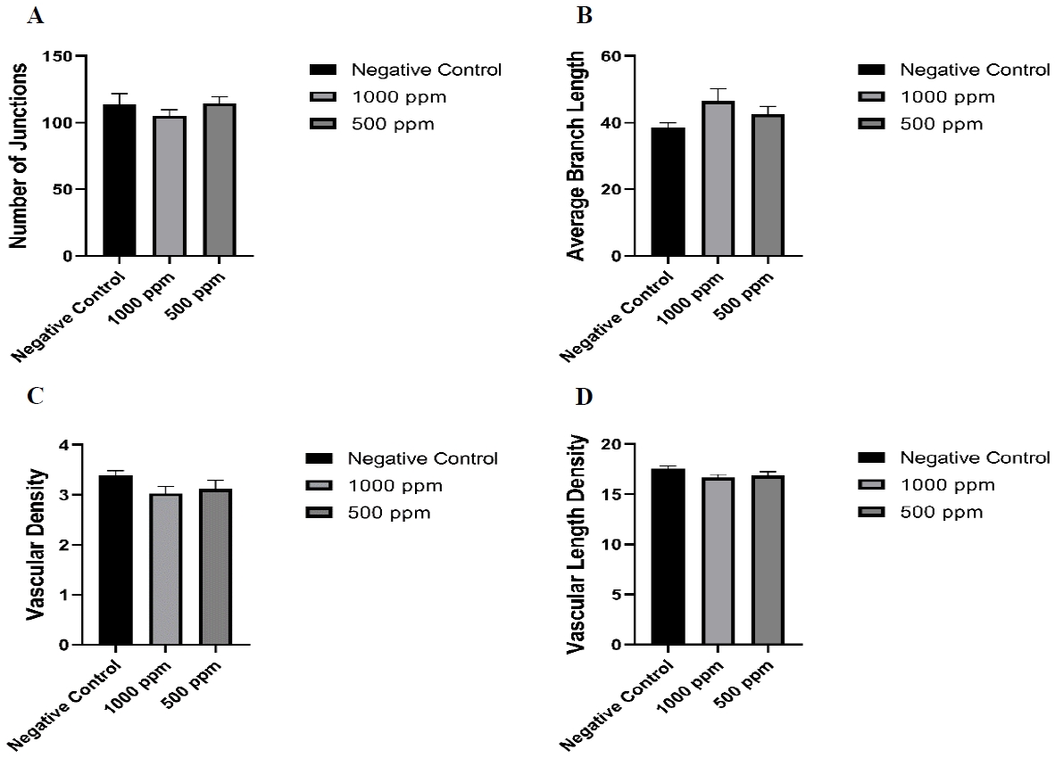
The generating steps of the ImageJ parameters from the captured images are presented in Fig. 2. At the time of image acquisition (day 7 of incubation), the embryos were equivalent to Hamburger-Hamilton stage 28 [60]. In the control embryos, a rich network of vitelline circulation was observed surrounding the embryo (Fig. 2A). While in the treated groups, there was an observable thinning of the veins, especially those treated with 1000 ppm of the ethyl acetate leaf extract (Fig. 2B). The number of junctions of vessels, vascular density and vascular length density were decreased as compared with the controls but not statistically significant (Supplementary (Fig. 3A, B; 3C, D).
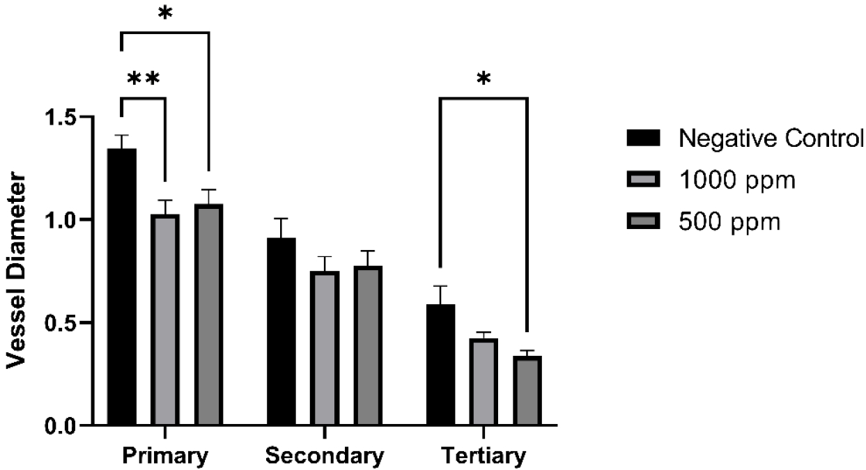
The CAM's vessel diameter distribution was found to be normal. However, the primary vessel diameters of the CAM treated with 1000 ppm and 500 ppm from the crude leaf extracts were significantly decreased than that of the control group (p<0.0001 and p<0.0372 respectively) (Fig. 4). It was also noted that the secondary and tertiary vessel diameters of the D. peltatus-treated groups were decreased compared to the control group but not statistically significant (Fig. 4).
The first indication of the toxicity of D. peltatus leaf extract in the duck embryos is the inhibition of early vascular development on the CAM. The number of junctions of the blood vessels of the embryo treated with the highest dose of D. peltatus showed to be lesser than that of the control, indicating that D. peltatus does have an inhibiting effect on the CAM vasculature. Embryos treated with 1000 ppm of D. peltatus showed the highest value of average branch length compared to the other groups. This result could indicate that the vasculature of the embryos treated with 1000 ppm of the extract contains long secondary branches and only a few tertiary vessels compared to the control and 500 ppm groups. The vascular density and vascular length density trends were almost identical. The ethyl acetate extract shows a reduction in vascularization in both measures. A comprehensive review by Olatunji et al. [61] on the genus Merremia, a homotypic synonym of Decalobanthus peltatus, shows that Merremia species have the potential to be developed into chemotherapeutic drugs since they demonstrated considerable cytotoxic effects and suppressed cancer cell growth. These results were attributed to different phytochemicals present in the species, including alkaloids, flavonoids, leucoanthocyanidins, steroids, and phenolics [21, 22, 61]. In addition to their ability to alter the activity of vital cellular enzymes, flavonoids are also crucial in medicine due to their anti-oxidative, anti-inflammatory, anti-mutagenic, and anti-carcinogenic properties [48]. Moreover, a studied alkaloid called sanguinarine from the roots of Sanguinaria canadensis has been reported to markedly suppress VEGF-induced endothelial cell migration and sprouting in vitro and blood vessel formation in vivo by blocking VEGF-induced Akt phosphorylation in a dose-dependent manner (10–300 nM) [62]. These bioactive compounds may also be involved in anti-vasculogenesis and anti-angiogenesis, which explain the reduction of the vascular plexus in injected embryos in the current investigation.
Gross morphological evaluation of the embryo
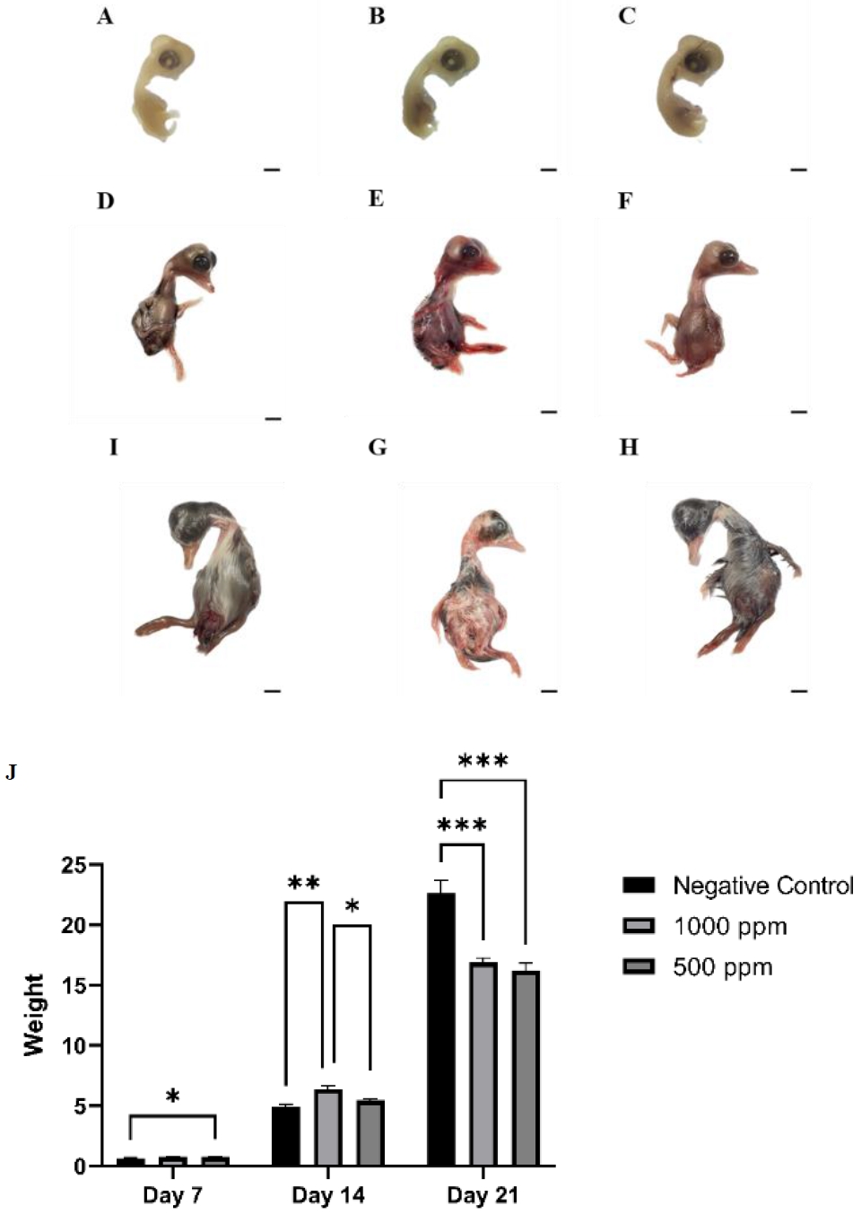
Gross morphological analysis of control and ethyl acetate leaf extract treated embryos during the first, second, and third trimesters of the growing period (days 7, 14, and 21 of the incubation periods, respectively) are shown in Figure 5. Embryos that are treated with both concentrations of D. peltatus showed no significant malformations in the first trimester (Fig. 5A, B, C). Gross evaluation of the embryos during the second trimester shows swelling and redness on the external body surfaces of both concentrations of the treated embryos compared to the control. The abnormalities are found mainly on the skin and appear as local or general hemorrhages (Fig. 5A, B, C). During the third trimester, more than half of the treated embryos show disorders characterized by moderate general redness and growth retardation (Fig. 5H, I). In comparison, all the control embryos and those that received a low extract concentration exhibited no gross disorder (Fig. 5A, D, and G).
The weight of the treated embryos at three different time points (days 7, 14, and 21 of the incubation period) are displayed in Figure 5J. The D. peltatus-treated embryos that received the 1000 ppm of D. peltatus were lighter as compared to the control (0.75 ± 0.02 g (95 % CI 0.70 ± 0.80), on day 7; 6.410 ± 0.25 g (95 % CI 5.86 ± 6.7), p =0.0010, on day 14; 16.88 ± 0.37g (95 % CI 16.04 ± 17.72), p < 0.0001, on day 21) (Fig. 5J). The embryos that received 500 ppm of plant extract showed a weight loss comparable to the control only at days 7 and 21 of the incubation period (0.78 ± 0.03 g (95 % CI 0.71 ± 0.85), p=0.0276 on day 7; 5.450 ± 0.13 g (95 % CI 5.15 ± 5.75), on day 14; 16.24 ± 0.61 g (95 % CI 14.86 ± 17.62), p < 0.0001on day 21) (Fig. 5J). The control embryos had average weight (0.66 ± 0.03 g (95 % CI 0.59 ± 0.73) on day 7; 4.92 ± 0.02 g (95 % CI 4.4 ± 5.4) on day 14; 22.66 ± 1.06 g (95 % CI 20.25 ± 25.07), and on day 21 (Fig. 5J).
Histopathological evaluation
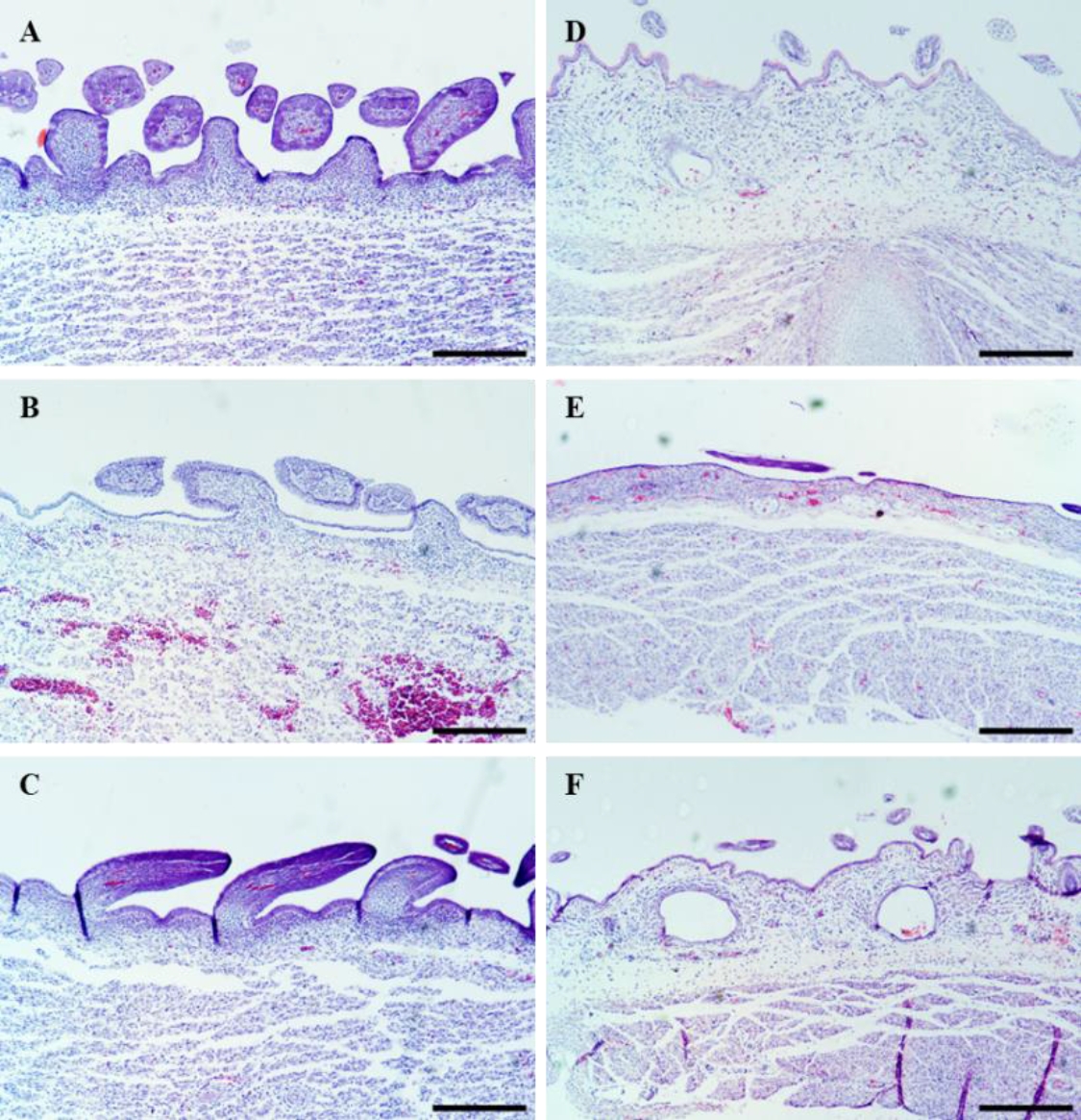
Skin tissues from control and treated duck embryos at different time points were subjected to histopathological analysis (Fig. 6). The negative control group had normal skin layer morphology, including a thick intact epidermal layer with apparent dermis and with the presence of blood vessels (Fig. 6).
On day 14 of the incubation period (11 days after treatment with 1000 ppm of D. peltatus extract), histopathological analysis showed a reduced number of feather follicles as well as damage to the blood vessels (Fig. 6B). While at 500 ppm, the skin of the embryo shows no signs of damage to its epidermis and underlying dermis (Fig. 6C).
At day 21 of the incubation period (19 days after treatment with 1000 ppm), sloughing of the epidermis was observed in all the samples as compared to control (Fig. 6E). The structure of the skin included abnormal dermis and hypodermis morphology with significant thinning of the epidermal layer and larger blood vessels were observed in 500 ppm crude ethyl acetate leaf extract as compared to the control (Fig. 6F).
The severity of the gross and histopathological abnormalities was highest in the embryos treated with the highest extract concentration. The toxic effect on the skin may be due to some of the bioactive compounds present in the extract. Several Merremia species of the Convolvulaceae family are toxic to ruminants. De Brito et al., for example, observed spontaneous Merremia macrocalyx (Ruiz & Pav.) O'Donell poisoning in cattle in Pernambuco state, north-eastern Brazil [63]. Furthermore, Adebiyi and colleagues conducted an in-depth study assessing the acute and chronic toxicity of an ethanolic leaf extract obtained from Merremia tridentata (L.) Hallier f. using albino male rats as the experimental subjects. Their findings indicated that chronic exposure to the plant extract at a dose of 100 mg/kg did not result in any significant deviations in biochemical parameters compared to the control group. However, when higher doses of 200 and 400 mg/kg were administered, a substantial elevation in biochemical markers, including creatine, urea, transaminases, and creatine kinase, was observed. These outcomes suggest a potential for adverse effects on the kidney, liver, and heart [64]. Phytochemical investigations of D. peltatus revealed the presence of alkaloids [21]. A study conducted by Zhao et al. (2011) suggests that the intake of a type of alkaloid called pyrrolizidine alkaloid is responsible for skin cancer since it can lead to photosensitization in animals upon their consumption and metabolism [65]. Although the specific concentration of leaf extracts used by locals for pathological treatments is unknown, it is crucial to highlight these findings, which imply that D. peltatus leaf extract at high concentrations can cause skin toxicity.
Conclusions
This study presents the extraction of D. peltatus leaves using various solvents. Among these solvents, ethyl acetate exhibited the highest toxicity, as evidenced by the lowest LC50 value. Duck embryos treated with the highest concentration of D. peltatus extract displayed malformations, growth retardation, and hemorrhages on their external body surfaces. While the specific concentrations of leaf extracts used for traditional medicinal purposes remain uncertain, these findings underscore the potential for skin toxicity associated with high concentrations of D. peltatus leaf extract. Therefore, comprehensive toxicity assessments, including herbal toxicokinetics and high-throughput genetic sequencing, should be conducted to ensure the safety of therapeutic herbs on embryos.